The Photochemical Machinery: Photoreceptor Bleaching and Retinal Physics
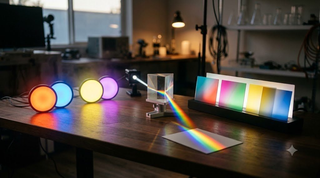
The sensation of color is not an inherent property of electromagnetic radiation; photons possess wavelengths and frequencies, but they do not possess hues. Color is a biological invention—a complex neurological code synthesized by the primate brain to discriminate surfaces, evaluate biological ripeness, and segment objects across volatile illumination conditions. The human retina achieves this sensory marvel through three distinct classes of cone photoreceptors, characterized by their peak spectral sensitivities: S-cones (short-wavelength, blue, peak ~420 nm), M-cones (medium-wavelength, green, peak ~530 nm), and L-cones (long-wavelength, red, peak ~560 nm).
However, the biochemical mechanisms that allow the retina to capture light are fundamentally constrained by metabolic and molecular physical limits. Photoreceptor signaling relies on phototransduction: when a photon is absorbed by an 11-cis-retinal chromophore bound to an opsin G-protein coupled receptor (rhodopsin in rods, photopsin in cones), the chromophore isomerizes into all-trans-retinal. This conformational shift triggers an intracellular biochemical cascade that hydrolyzes cyclic guanosine monophosphate (cGMP), closing sodium/calcium ion channels and hyperpolarizing the photoreceptor membrane. This event reduces the tonic release of the inhibitory neurotransmitter glutamate onto downstream bipolar cells.
Because the enzymatic regeneration of 11-cis-retinal via the retinal pigment epithelium is a finite metabolic process, prolonged exposure to intense, unvarying visual stimuli exhausts the available photopigment pool—a state known as ‘photopigment bleaching’. This physiological exhaustion forms the biological engine of physiological optical illusions: negative afterimages, Troxler fading, Mach bands, and dramatic simultaneous contrast shifts. In this comprehensive study, we explore the cellular biophysics, opponent-process genetics, and cortical color constancy algorithms that shape our chromatic reality.
🧬 Key Biological Concept: The Retinal Pigment Epithelium (RPE)
All-trans-retinal cannot be re-isomerized inside the photoreceptor outer segment. It must be actively transported into the adjacent retinal pigment epithelium, biochemically converted back into 11-cis-retinal through the RPE65 isomerase enzyme, and shuttled back to the photoreceptor. This cycle takes tens of seconds, creating the temporal window for afterimages.
Troxler’s Fading: Why the Brain Erases Static Reality
In 1804, Swiss physician and philosopher Ignaz Paul Vital Troxler made an extraordinary discovery: if an observer fixes their unwavering gaze upon a stationary central focal point, stationary visual stimuli positioned in the peripheral visual field begin to fade, dissolve, and completely vanish within twenty to thirty seconds, leaving behind only the solid color of the background. The most famous modern demonstration of this phenomenon is the ‘Lilac Chaser’ or ‘Pac-Man’ illusion, where a circle of blurred lilac dots fades entirely until a lone green dot appears to run smoothly around the circular track.
Troxler’s fading reveals a cardinal operating principle of sensory neurobiology: the nervous system is tuned to detect change, novelty, and temporal differentials, not invariant continuity. At the level of the retina and the lateral geniculate nucleus (LGN), neural receptive fields respond vigorously to luminance and chromatic transients (temporal derivatives). If an image remains completely immobile across the exact same patch of retinal photoreceptors, the biochemical transduction cascade reaches a static chemical equilibrium. Intracellular calcium feedback diminishes guanylyl cyclase activity, arrestin quenches activated rhodopsin, and downstream ganglion cells cease firing action potentials.
Why, then, does our entire visual world not fade into darkness during normal wakefulness? The answer lies in the constant, microscopic motor activity of the human eye. Even during our most concentrated attempts at absolute visual fixation, our ocular muscles execute three distinct categories of microscopic movements: low-amplitude high-frequency physiological tremor (tremor), slow wandering ocular drifts, and rapid corrective flicks termed microsaccades. These micro-movements ensure that high-contrast boundaries are constantly swept across adjacent photoreceptors every few milliseconds, refreshing neural stimulation and preventing perceptual adaptation.
Why the Periphery Fades First: Receptive Field Scaling
Troxler fading occurs dramatically in the peripheral retina while sparing the fovea because of dramatic differences in neural anatomy. In the central fovea, photoreceptor cones are microscopically tiny and connect in a 1:1 ratio with midget bipolar and midget ganglion cells, yielding minuscule receptive fields capable of detecting microscopic tremors. In the periphery, conversely, hundreds of photoreceptors converge onto single parasol ganglion cells, producing massive receptive fields. Micro-ocular drifts are insufficient to shift an edge outside these expansive peripheral receptive fields, allowing peripheral neural adaptation to proceed unchecked, leading to total perceptual erasure.
| Physiological Illusion | Discoverer / Pioneer | Retinal / Cortical Locus | Key Biochemical / Neural Mechanism | Perceptual Manifestation |
|---|---|---|---|---|
| Negative Afterimages | Jan Evangelista Purkyně (1819) | Retinal cones & Hering opponent channels | Photopigment bleaching & opponent rebound firing | Complementary color phantom seen on white neutral card |
| Troxler Fading | Ignaz Paul Vital Troxler (1804) | Peripheral retinal ganglion cells & LGN | Neural adaptation & lack of saccadic edge refreshment | Peripheral stationary objects dissolve into background |
| Simultaneous Contrast | Michel Eugène Chevreul (1839) | Retinal ON/OFF bipolar cells & V1 | Horizontal cell lateral inhibition | Identical grey swatch appears darker on light background |
| Mach Bands | Ernst Mach (1865) | Retinal horizontal cell interneurons | Spatial second derivative of luminance gradient | Exaggerated light/dark banding at luminance boundaries |
| Land’s Retinex Effect | Edwin H. Land (1959) | Cortical visual area V4 | Global spatial ratio comparison across visual fields | Color constancy maintained under extreme chromatic shifts |
| The McCollough Effect | Celeste McCollough (1965) | Binocular orientation units in V1 | Long-term associative plastic neural habituation | Contingent color aftereffects lasting days or weeks |
Hering’s Opponent-Process Theory and Negative Afterimages
If you stare fixedly at a bright, saturated red square for forty seconds and then immediately glance at a clean, neutral white wall, an unmistakable, ghostly cyan square appears hovering before your eyes. If you stare at a saturated yellow pattern, its negative afterimage emerges in vivid blue; a vibrant green target produces an afterimage of rich magenta. This complementary chromatic pairing is direct behavioral proof of Ewald Hering’s Opponent-Process Theory of color vision.
While the Young-Helmholtz trichromatic theory correctly describes color reception at the photoreceptor layer (red, green, and blue cones), Hering’s opponent framework accurately models how those cone signals are organized and processed by downstream retinal ganglion cells and neurons in the lateral geniculate nucleus (LGN). The visual system organizes color into three antagonistic, mutually inhibitory channels:
- The Red-Green Channel (L vs. M cones): Excitation by red suppresses green; excitation by green suppresses red.
- The Blue-Yellow Channel (S vs. L+M cones): Excitation by blue suppresses yellow; excitation by yellow suppresses blue.
- The Achromatic Luminance Channel (L + M + S cones): Encodes lightness versus darkness (black vs. white).
When you fixate upon a saturated green stimulus, the green-selective input units in the red-green opponent channel are driven to their maximum metabolic output, while the red-selective units are held in deep synaptic inhibition. Over dozens of seconds, the green cones experience profound photopigment bleaching, and their downstream synapses adapt. When you shift your gaze to a neutral white surface—which emits an equal distribution of all visible wavelengths—the exhausted green pathway fails to fire with normal vigor. The rested, unadapted red pathway, suddenly released from inhibition, responds with robust spontaneous baseline activity. The opponent comparator decodes this massive neural imbalance as red/magenta light, generating a vivid negative afterimage that endures until the photopigments regenerate.
The McCollough Effect: An Aftereffect That Endures for Weeks
Discovered by American psychologist Celeste McCollough in 1965, the McCollough Effect stands as one of the most enigmatic phenomena in perceptual science. In this experiment, an observer alternates between gazing at red horizontal stripes and green vertical stripes for ten to fifteen minutes. Afterwards, when the subject looks at black-and-white test grids, the horizontal black-and-white lines appear distinctly greenish, while the vertical black-and-white lines appear distinctly pinkish or reddish.
Unlike standard photochemical afterimages that fade within twenty to thirty seconds as retinal photopigments regenerate, the McCollough Effect is orientation-contingent and possesses astonishing longevity: it can persist for days, weeks, and in some documented experimental cases, over three months! Because retinal cones have no orientation selectivity—orientation tuning first emerges in cortical area V1—the McCollough Effect proves that neural adaptation can occur at the level of cortical synapses. It represents a form of associative classical conditioning or long-term synaptic depression within the primary visual cortex, demonstrating that visual exposure can physically reconfigure cortical processing pathways for extended periods.
Simultaneous Contrast and Chevreul’s Laws
In 1839, French chemist and director of the Gobelins Royal Tapestry Works, Michel Eugène Chevreul, published ‘The Law of Simultaneous Contrast of Colors’. The tapestry weavers were being accused of using substandard, faded dyes because certain woven threads appeared dull and washed out in finished carpets. Chevreul’s scientific investigation revealed that the chemical composition of the dyes was impeccable; the problem was purely physiological. When two threads of contrasting hues or lightness values were woven immediately adjacent to one another, each color radically modified the perceived appearance of its neighbor.
Simultaneous contrast is governed by lateral inhibition mediated by horizontal cells in the outer plexiform layer of the retina. Horizontal cells form an extensive, electrically coupled syncytium (via gap junctions) that stretches laterally across the photoreceptor mosaic. When a photoreceptor is strongly hyperpolarized by bright light, it signals adjacent horizontal cells, which in turn feed back inhibitory signals onto neighboring photoreceptors.
As a direct consequence of this lateral inhibition, a medium-gray square situated inside an expansive black surround appears brilliantly luminous, crisp, and pale; the identical medium-gray square placed inside a pure white surround appears dingy, murky, and nearly black. The white surround vigorously activates horizontal cells, which cast an inhibitory blanket over the central gray region, depressing its neural output. The black surround, generating minimal horizontal cell feedback, allows the central gray region to fire uninhibited. The brain does not evaluate absolute photon counts; it measures contrast ratios against local environmental surroundings.
Mach Bands: The Mathematics of Edge Sharpening
Discovered in 1865 by Austrian physicist Ernst Mach, Mach Bands are illusory bright and dark bands that appear at the boundaries of a continuous luminance gradient. When a visual surface transitions smoothly from a uniform dark region through a ramp gradient to a uniform light region, the human eye perceives an exaggerated bright stripe just where the ramp meets the light area, and an exaggerated dark line where the ramp meets the dark area. In physical reality, optical photometers show no such peaks or troughs in luminance; the gradient is strictly monotonic.
Mach mathematically demonstrated that this phenomenon represents the biological computation of the spatial second derivative of luminance. By applying lateral inhibition across spatial receptive fields, the retina acts as a Laplace operator, exaggerating inflection points where the rate of change of luminance alters. This physiological mechanism is vital for biological survival: it sharpens blurry contours in low-light environments, enabling animals to detect the subtle silhouettes of concealed predators against complex foliage.
🎨 Chevreul’s Practical Dictum for Visual Artists
‘Two complementary colors, placed side-by-side, each acquire a heightened degree of brilliant saturation; whereas two non-complementary colors mutually corrupt each other’s chromatic integrity.’ This rule founded the Post-Impressionist Divisionist and Pointillist movements championed by Georges Seurat.
The Miracle of Color Constancy: Edwin Land and the Retinex Theory
One of the most profound evolutionary feats of the human visual system is ‘Color Constancy’—the ability to perceive a green apple as consistently green whether viewed under the cool, bluish illumination of outdoor twilight (which contains heavy short wavelengths) or under the warm, amber glow of an indoor incandescent tungsten bulb (which is saturated with long red wavelengths). A standard camera sensor reading raw RGB values would register two entirely different chromatic objects; the human visual cortex, however, perceives the apple as having an identical surface reflectance.
In 1959, the brilliant inventor of the Polaroid camera, Edwin H. Land, conducted a series of revolutionary experiments that shattered classical optics. Land projected complex abstract collages of colored papers (‘Mondrian’ displays) using three separate black-and-white slide projectors equipped with red, green, and blue color filters. Land carefully calibrated the light output such that a specific green patch in the Mondrian display reflected the exact same radiometric spectrum of light (identical milliwatts per wavelength) as a nearby red patch had reflected in a previous trial.
Under classical Newtonian optics, the patch should have looked identical. In reality, human observers continued to perceive the green patch as vividly green, and the red patch as unmistakably red! Land concluded that color cannot be computed locally from the wavelength reaching the eye from an isolated point. Instead, the brain calculates global spatial ratios across the entire visual field. Land formulated the ‘Retinex Theory’ (a portmanteau of retina and cortex), proposing that the visual cortex possesses three independent lightness records across short, medium, and long wavelengths, and compares the relative ratios between adjacent surfaces to discount the ambient illuminant.
Cortical Area V4: The Seat of Chromatic Constancy
Modern functional neuroimaging and electrophysiological recordings by Semir Zeki and colleagues have confirmed that Land’s Retinex computations take place in visual cortical area V4 within the lingual and fusiform gyri. While neurons in primary visual cortex (V1) fire strictly in response to the physical wavelength of light entering their narrow receptive fields (acting like simple spectrometers), neurons in area V4 possess expansive receptive fields that integrate signals across the entire visual scene. V4 neurons maintain constant firing rates for specific perceived colors regardless of dramatic shifts in the spectral composition of the illuminating light source. When area V4 is damaged by ischemic stroke or trauma, patients develop cerebral achromatopsia—a tragic condition where the physical eyes remain intact, yet the conscious world is rendered entirely in drained shades of gray, stripped of the vibrant subjective dimension of chromatic experience.
Conclusion: The Deconstructed Eye
Physiological optical illusions demonstrate that our eyes are biological instruments subject to the laws of physical chemistry, enzymatic kinetics, and cellular fatigue. Yet out of these physical constraints emerges an astonishing computational architecture. By sacrificing static accuracy through Troxler fading, our visual system conserves immense metabolic energy. By deploying lateral inhibition and Chevreul contrast, the retina sharpens edges and detects camouflage. And through the complex cortical algorithms of area V4, our brain constructs a stable, color-constant world from an ever-shifting sea of ambient photons.