The Dynamic Eye: How the Brain Computes Velocity and Motion
Motion perception is among the most ancient and vital functions of the vertebrate visual apparatus. Long before organisms developed high-acuity color vision or the complex cortical machinery necessary for fine facial recognition, they required the immediate capacity to detect moving contours against static backgrounds to evade predators and capture prey. In the human brain, visual motion processing is handled by a specialized neural hierarchy anchored in the Middle Temporal visual area (often designated as area MT or V5), which receives rapid, low-latency magnocellular signals directly from the primary visual cortex (V1) and the superior colliculus.
However, despite the extraordinary temporal precision of area MT, motion perception is remarkably susceptible to physiological adaptation, neural fatigue, and spatial contrast exploitation. When an observer gazes fixedly at continuous, unidirectional visual motion for an extended duration, the cessation of that motion reveals a stunning perceptual anomaly: static objects appear to drift, crawl, and expand in the diametrically opposite direction. This phenomenon, universally known as the Motion Aftereffect (MAE) or the Waterfall Illusion, provides definitive empirical evidence that motion perception is governed by an antagonistic, opponent-process neural architecture.
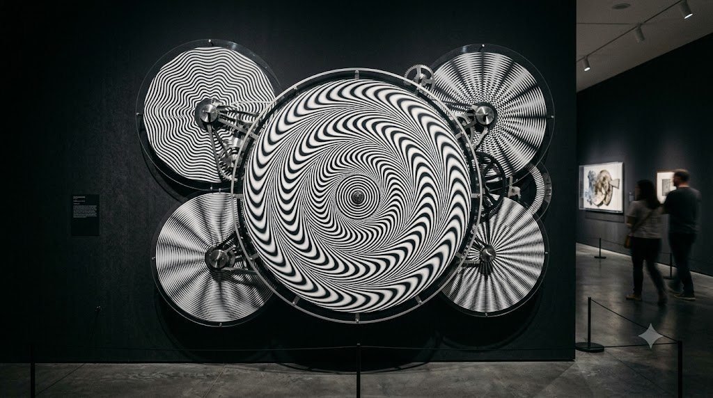
In parallel, the mid-twentieth century witnessed the rise of Op Art (Optical Art)—an avant-garde movement pioneered by visionaries such as Bridget Riley, Victor Vasarely, and later neuroscientist Akiyoshi Kitaoka. These artists and researchers unlocked static geometric configurations that stimulate retinal transients and microsaccadic eye movements, tricking the visual cortex into perceiving vigorous illusory motion across completely stationary canvases. This deep analytical treatise explores the neurophysiology of motion aftereffects, cortical motion tuning, and the exquisite mechanics of peripheral drift illusions.
🌊 Historical Milestone: Aristotle and the Running Stream
The earliest documented description of the Motion Aftereffect dates back to Aristotle’s treatise ‘Parva Naturalia’ (circa 350 BCE), where he noted that after observing a rushing river, glancing at stationary stones on the bank made the pebbles appear to move in reverse. In 1834, Scottish scientist Robert Addams formalized the experiment at the Falls of Foyers, establishing the Waterfall Illusion.
Neurobiology of the Motion Aftereffect: Cortical Population Vectors
To understand why static surfaces drift backward following motion adaptation, one must examine the receptive field dynamics of direction-selective neurons within cortical area V5/MT. Neurons in this specialized region exhibit precise directional tuning: an individual MT neuron will fire with maximum frequency when visual stimuli traverse its receptive field in its ‘preferred’ direction (for example, downward at 5 degrees per second), whereas stimuli moving in the opposite ‘null’ direction produce complete baseline inhibition.
Under normal conditions, when an individual gazes upon a completely static scene, directionally tuned neurons across all vectors (up, down, left, right, diagonals) discharge at an identical, baseline spontaneous firing rate (typically between 5 to 10 action potentials per second). The visual cortex decodes motion through population vector coding: it calculates the vector sum of all directional outputs across the population. Because the spontaneous baseline discharges in opposing directions cancel each other out symmetrically, the resultant vector magnitude is zero, and the scene is consciously perceived as stationary.
However, when an observer stares intently at a cascading waterfall for 30 to 60 seconds, downward-tuned MT neurons fire at their maximum physiological threshold. This sustained metabolic exertion leads to profound neural adaptation, caused by the depletion of presynaptic neurotransmitter vesicles, hyperpolarization of the postsynaptic membrane via calcium-activated potassium channels, and active synaptic inhibition. When the observer abruptly shifts their gaze to the stationary rock face adjacent to the falls, downward-tuned neurons are so thoroughly exhausted that their firing rate collapses well below their baseline resting rate.
Crucially, the upward-tuned opponent neurons—having experienced no excitation during the adaptation phase—continue firing at their normal, spontaneous baseline rate. When the visual cortex calculates the population vector sum of this post-adaptation state, the upward signal significantly outweighs the suppressed downward signal. The resulting net neural vector points directly upward, creating the undeniable perceptual hallucination that the solid rock wall is ascending toward the sky. The illusion endures until metabolic homeostasis is restored and presynaptic vesicles are replenished, usually lasting between 10 to 30 seconds.
Interocular Transfer: Proving the Cortical Nature of MAE
One of the most profound psychophysical demonstrations in vision science is the phenomenon of interocular transfer. If an observer covers their left eye completely with an opaque patch and adapts their right eye to downward motion on a screen for sixty seconds, and then quickly swaps the patch so that the unadapted left eye views a static test pattern, the observer still perceives a robust upward motion aftereffect. Because signals from the two eyes remain anatomically segregated throughout the retina, the optic nerve, and the lateral geniculate nucleus (LGN)—only combining binocularly within layer IV of primary visual cortex (V1) and subsequent associative areas like V5/MT—interocular transfer provides conclusive proof that the motion aftereffect does not originate from retinal fatigue, but is a high-level cortical calculation.
Furthermore, functional imaging reveals that the velocity and duration of the perceived aftereffect correlate precisely with the duration of the adaptation phase, obeying logarithmic decay curves. When adaptation is prolonged over several minutes, the perceptual aftereffect can persist for hours, a phenomenon known as the ‘long-lasting motion aftereffect’, indicating that sustained directional exposure recruits plastic synaptic modifications akin to long-term depression (LTD) within cortical circuits.
| Motion Illusion Class | Key Paradigm / Artifact | Primary Cortical Area | Underlying Neural Mechanism | Diagnostic Diagnostic Trait |
|---|---|---|---|---|
| Classical Motion Aftereffect | Waterfall Illusion / Addams (1834) | Area V5/MT & V1 direction units | Opponent neural fatigue & population vector imbalance | Reverse drift on static surfaces following adaptation |
| Interocular Transfer MAE | Dichoptic Motion Adaptation | Binocular neurons in V1 & MT | Transfer of adaptation from adapted eye to unadapted eye | Proves cortical rather than retinal locus of adaptation |
| Peripheral Drift Illusion | Rotating Snakes (Kitaoka, 2003) | V1/V2 contrast latency differences | Asymmetric luminance steps & microsaccadic triggers | Perceived circular rotation in peripheral visual fields |
| Enigma Illusion | Isia Léviant (1981) | Macula & early retinotopic cortex | Microsaccadic jitter over concentric high-frequency rings | Rapid shimmering streaming inside color rings |
| Motion-Induced Blindness | Bonneh, Cooperman, Sagi (2001) | Ventral stream & parietal attentional gating | Attentional competition between global flow and static targets | Spontaneous total disappearance of bright salient yellow dots |
| The Barberpole Effect | Wallach (1935) | V1 end-stopped cells & MT aperture integration | Aperture problem resolving along elongated boundary edges | Diagonal stripes perceived moving strictly up or down |
Op Art and the Science of Static Kinetic Illusion
During the 1960s, the international art world was revolutionized by the emergence of Optical Art, led by British artist Bridget Riley. In iconic masterworks such as ‘Movement in Squares’ (1961) and ‘Current’ (1964), Riley demonstrated that precise geometric draftsmanship could evoke violent sensations of undulating movement, shimmering vibrations, and three-dimensional spatial warping without incorporating a single motor or moving part.
Riley’s compositions exploit the spatial frequency tuning of cortical orientation columns. When high-contrast black and white lines are drawn with mathematically compressed spatial intervals, they approach the resolving threshold of human foveal vision. At these critical frequencies, normal physiological ocular tremor—involuntary micro-movements of the eye occurring at 30 to 70 Hz, known as tremor, drifts, and microsaccades—causes the edges of the high-contrast lines to sweep across adjacent photoreceptors with extreme speed. The visual cortex misinterprets these microscopic retinal shifts as macroscopic kinetic flow, generating an irresistible sensation of surging waves.
Akiyoshi Kitaoka and the Rotating Snakes Phenomenon
In 2003, Japanese vision scientist and psychologist Akiyoshi Kitaoka unveiled what is widely regarded as the pinnacle of static motion illusions: the ‘Rotating Snakes’. This mesmerizing image features an array of circular patterns composed of repeating step-wise color sequences: typically black, dark blue, white, and light yellow. When an observer scans across the image, the circular ‘snakes’ appear to rotate vigorously in the peripheral visual field, yet freeze into immobility the instant the observer fixes their direct foveal gaze onto any individual circle.
The neurophysiological mechanism responsible for the Rotating Snakes illusion is the Peripheral Drift Illusion (PDI), which relies on luminance contrast latency differences in early visual processing. In the mammalian retina, visual signals generated by high-contrast edges (such as pure black transitioning to pure white) are processed and transmitted through the optic nerve significantly faster than signals generated by low-contrast boundaries (such as dark blue transitioning to black, or light yellow transitioning to white). This latency differential spans approximately 10 to 40 milliseconds.
When the human eye executes a natural saccade or involuntary microsaccade across the pattern, the retinal receptors are stimulated simultaneously across all sectors of the circular band. However, because of the contrast-dependent latency discrepancy, the high-contrast neural signals arrive at the visual cortex first, followed sequentially by intermediate and low-contrast signals. The motion-sensitive direction-selective neurons in area MT interpret this temporal arrival delay across spatially adjacent steps as physical directional motion. As long as the eye continues making micro-exploratory movements across the canvas, the circular snakes appear to spin endlessly.
🎯 Laboratory Verification: The Stabilization Paradigm
When the ‘Rotating Snakes’ image is stabilized on the retina using specialized eye-tracking optical apparatus that moves the display in absolute sync with ocular tremor, the illusory motion completely and instantaneously ceases. This confirms that involuntary eye movements are the essential physical spark that ignites peripheral drift.
The Enigma Illusion: Microsaccades and Cortical Shimmering
In 1981, French painter Isia Léviant created ‘Enigma’, an abstract painting consisting of radiating black and white spokes overlaid with several concentric rings of solid, untextured color (such as purple, ochre, or green). When observers look at the center of Enigma, a rapid, rotary streaming or pulsating current of rapid motion is perceived circulating furiously through the colored rings, resembling fluid rushing through a transparent circular conduit.
For decades, neuroscientists argued whether Enigma’s shimmering motion was caused by rapid accommodative changes in the lens of the eye or by higher-level cortical processing. In a definitive study led by neurobiologist Susana Martinez-Conde and Stephen Macknik at the Barrow Neurological Institute, researchers used ultra-high-speed precision eye tracking synchronized with functional neuroimaging. They demonstrated that the subjective perception of motion within the Enigma rings correlates with microsaccadic frequency. When subjects executed a burst of microsaccades, the illusory streaming accelerated; when microsaccades were suppressed, the streaming slowed or halted. The radiating black spokes generate high-contrast square-wave luminance transients that stimulate magnocellular pathways in primary visual cortex, transforming tiny eye twitches into continuous subjective motion.
Motion-Induced Blindness: The Attentional Erasure of Salient Reality
First formally documented by Yoram Bonneh, Alexander Cooperman, and Dov Sagi in 2001, Motion-Induced Blindness (MIB) is a striking perceptual phenomenon that demonstrates how dominant kinetic signals can actively extinguish conscious awareness of completely static visual objects. In a standard MIB setup, an observer fixates on a central flashing green cross while a cloud of brilliant blue dots rotates continuously in the background. Stationed within this moving field are three bright, high-contrast, stationary yellow dots arranged in a triangle.
Within seconds of maintaining central fixation, the brilliant yellow dots begin to disappear from conscious perception—either individually or simultaneously—for durations spanning several seconds at a time, before reappearing with startling clarity. The physical photons from the yellow dots never cease striking the retinal photoreceptors, yet the visual cortex actively deletes them from the perceived mental image.
Neuroscientists attribute MIB to an evolutionary mechanism known as perceptual filling-in and surface completion. In natural vision, stationary items that fall within a coherent, moving global flow field are often interpreted by higher-order parietal attentional networks as visual artifacts, optical debris, or retinal scotomas (blind spots). To construct a clean, continuous perceptual scene, the visual cortex suppresses the neural representation of the static targets in favor of the coherent global motion field, demonstrating that what we see is governed as much by attentional filtering and suppression as it is by incoming physical light.
The Barberpole Illusion and the Aperture Problem
One of the classic puzzles of visual neuroscience is the ‘Aperture Problem’, brilliantly illustrated by the classic Barberpole illusion. A traditional barberpole consists of a vertical rotating cylinder painted with alternating diagonal red and white helical stripes. When the cylinder rotates horizontally on its vertical axis, observers perceive the stripes as traveling smoothly upwards (or downwards, depending on the rotation direction), rather than moving sideways.
The aperture problem arises because any individual motion-sensitive neuron in area V1 sees the world through a receptive field that acts as a microscopic aperture. If a straight, unoriented line segment moves across this aperture, the neuron can only measure motion perpendicular (orthogonal) to the line’s orientation; it is mathematically blind to any motion parallel to the line. Therefore, a diagonal stripe moving horizontally produces the exact same local velocity vector as a diagonal stripe moving vertically.
To resolve this profound ambiguity, the visual system relies on specialized ‘end-stopped’ neurons that detect the terminators—the physical points where the diagonal stripes intersect the borders of the aperture. In a vertical cylinder, there are far more terminators along the elongated vertical edges than at the short top and bottom ends. These edge terminators travel strictly in a vertical direction. Area MT integrates the ambiguous 1D contour signals with the unambiguous 2D terminator signals, allowing the vertical signals to capture the entire percept, demonstrating how global boundary constraints dictate our perception of local kinetic events.
Conclusion: The Evolutionary Imperative of Motion Illusions
Far from representing flawed sensory design, motion aftereffects and kinetic optical illusions demonstrate the remarkable efficiency of biological vision. By employing opponent-process coding, the visual system maximizes its dynamic sensitivity while minimizing metabolic energy expenditure. By leveraging latency differences to highlight rapid contrast transitions, the brain ensures immediate detection of predatory threats in peripheral fields.
Through the combined genius of psychophysicists, neuroscientists, and Op Art masters, we have learned to reverse-engineer these magnificent evolutionary adaptations. When Bridget Riley’s lines vibrate, when Kitaoka’s snakes slither across a motionless page, and when stationary cliffs soar into the sky after gazing upon Foyers Falls, we are witnessing the living machinery of the brain actively painting motion onto the canvas of human consciousness.